
Numbed and
Bared:
Intraoperative
Brain Mapping
by Emjay Rosales
No two brains are anatomically alike.
The function and the anatomy of our brain do not match a hundred percent. With this, such brain abnormalities alter the usual location of our biological functions. And when the abnormal mass of tissue or tumor is too close to some of the areas of the brain that are in charge of vision, body movements, and language, a certain kind of surgery is necessary to perform. This is called the awake craniotomy or intraoperative brain mapping.
WHAT IS INTRAOPERATIVE BRAIN MAPPING?
Intraoperative brain mapping is a brain surgery performed while the patient is awake. It is performed to treat epileptic seizures—a condition when there is a sudden surge of electrical activity in the brain—and brain tumors. The objective is to perform tumor removal while preserving the neurological function of the patient and without damaging it.
The biological functions can be seen in our four different lobes or the subdivision of the brain: the parietal lobe is in charge of sensory information including touch, pain, temperature, pressure; the frontal lobe is responsible for intellectual functions such as reasoning and emotional regulation; the temporal lobe is dedicated to process primary auditory complex or those of hearing and forming memories, and; the occipital lobe controls visual information from the eyes. Damage to these vital areas of the brain is minimized if the patient is awake during the surgery.

HOW IS INTRAOPERATIVE BRAIN MAPPING PERFORMED?
Prior to the surgery, the patient will be subjected firstly to anesthesia—a temporary loss of sensation or awareness through the use of injection, gas, or hypnosis before a medical procedure. The patient will receive nerve or scalp block—medicine injection to block the pain—to numb the scalp. The most popular agents for sedation or to produce tranquil state to the patient include propofol and dexmedetomidine. Propofol is sedative agent injected to the veins to induce and maintain anesthesia. Dexmedetomidine is provided without interfering respiration function or respiratory depression called hypoventilation.
The patient’s head is lying in a still position for a more accurate surgery procedure. The patient is asleep while the neurosurgeon is making an incision to his scalp and removing a certain area of the skull.
When the brain is finally exposed, neuroanaesthesiologists will stop the sedation and remove the patient’s breathing tube. The patient will then wakes up and participate during the critical part which is the mapping. During this moment, the patient will not feel any pain. This is because the brain has no pain receptors.
The neuropsychologist or the speech pathologist will start to ask the patient some questions, flash photos

in order to find a certain part of the brain to approach the lesion and try to take it out as safe as possible. The patient can be asked about things like "what is the color of the sky" or "name the months in a year." An abnormal change of tissue or organ that has been damaged from an injury or disease is called lesion.
WHAT IT FEELS LIKE

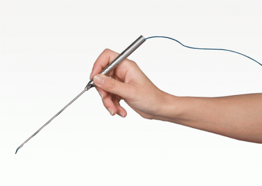
While this activity goes on, the neurosurgeon will make a temporary stun to a part of the brain using a small electrical probe—a slender medical instrument with a blunt end used to explore an opening or wound. There can be reactions that patients might find amusing and funny because of the stimulation induced in the areas responsible for such responses.
When current is applied, the patient may be triggered to laugh and giggle, or feel his foot rising without exerting an effort to. In the other moment, the stunned area where the patient momentarily stuttered or forgot to process information must be critically marked for it is important to avoid. This enables the surgeon to create a map of functions. Through this activity, the surgeon can pinpoint the safest neurological network to the affected area of the brain where the tumor resides.
WHAT IT LOOKS LIKE
The functional magnetic resonance imaging or fMRI is used in the procedure to see what is going on inside the brain in which there is a utilization of magnetic fields, centering on the brain’s metabolic function.
Neurosurgeon and Awake Craniotomy Specialist Ramin Rak, M.D., F.A.A.N.S, of NSPC described that brain mapping is performed for a reason.

“There’s always this concept of brain plasticity—the difference between everybody’s function location on the brain as opposed to their anatomy. So like when you do MRI and stuffs and so you see the anatomy and you know where things are, but again each patient’s function may be different in relation to anatomy and what we know,” said Rak.
The idea of an awake surgery might be frightening for some people. But there is an assurance that this kind of surgery requires advanced equipments and professionals, thus science got the patient’s back in the maximum utilization and safety performance of the procedure.